Degenerative Disc Disease
Pathology, Cause and Management

Intervertebral Disc
Function
The intervertebral disc makes up one fourth of the spinal column and can be described as pads of fibrocartilage-based structures found between the vertebral bodies within the spine (Donnally and Hanna, 2023). They connect the vertebrae and provide spinal mobility during particular movements whilst flexing forward and rotating the spine (Alomari et al., 2009). They provide support, flexibility and minor load-sharing (Donnally and Hanna, 2023). Vertebral discs act as a shock absorber between vertebrae and help mechanical stresses to be transmitted throughout the spine during movement from the lower limbs all the way up into the neck and skull (Alomari et al., 2009).
Disc Anatomy
The intervertebral disc is composed of two parts; an outer strong ring called the annulus fibrosus that surrounds a soft gel-like inner substance called the nucleus pulposus consisting of 80% to 85% water in normal/ non pathological cases.
Degenerative Disc Disease
Degenerative disc disease (DDD) is not a disease but a condition within the intervertebral disc.
Most intervertebral disc degenerations are asymptomatic with that patients can have the problem diagnosed (from scans) but not experience pain. This can make it difficult for physical therapy practitioners to diagnose and manage without the relevant medical imaging e.g. Magnetic Resonance Imaging (MRI).
Back pain symptoms related to intervertebral disc degeneration are caused by pathological changes with mainly being disc desiccation or a 'drying out' of the water content within a disc. The condition causes the following:
- Fibrosis; a thickening or scarring of tissue.
- Disc narrowing causing a reduction of disc height and/ or width.
- Can cause the annulus to bulge, fissure or undergo degeneration.
- Arthritis within the vertebrae.
Can This Create Nerve Irritation?
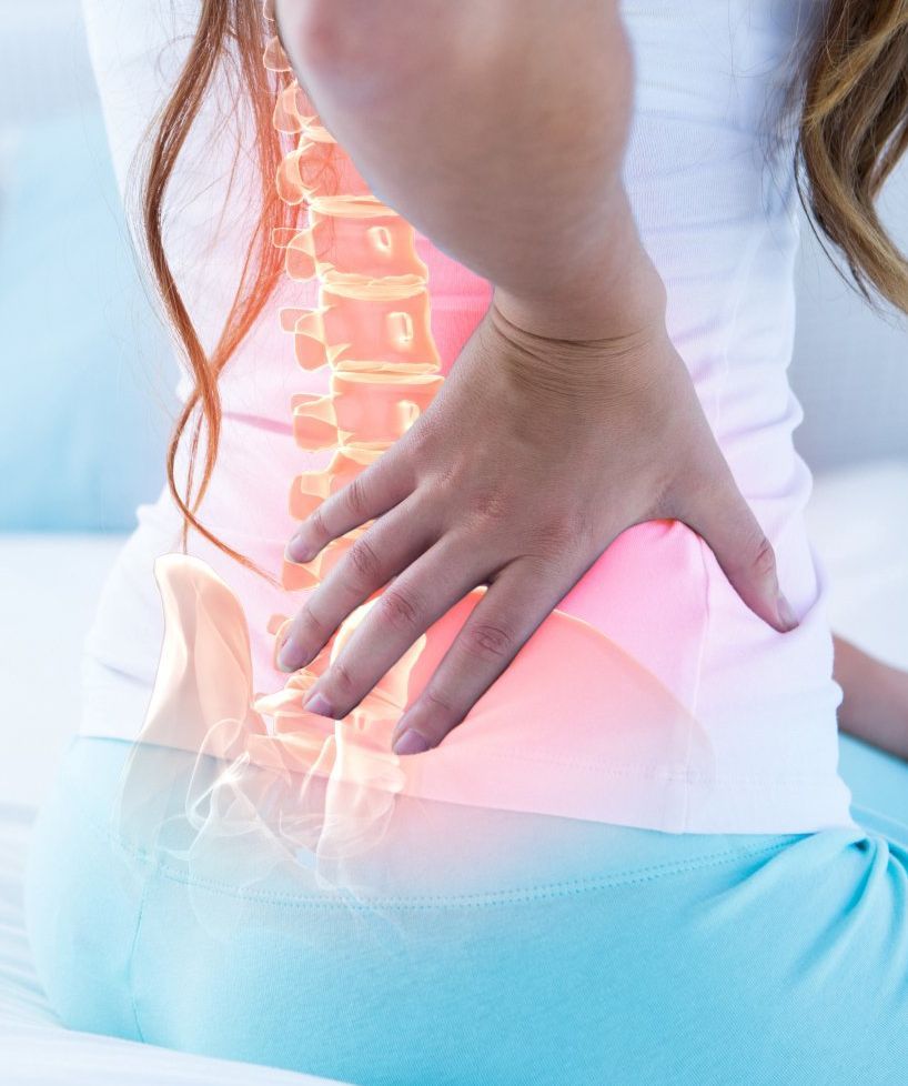
Progression can cause the disc to bulge and create pressure and irritation to neighbouring spinal nerves. This can lead to individuals experiencing tingling (paraethesia) into the limbs of the body such as the arms and fingers for if the region is affecting areas of the neck or buttock, thigh or toes for if occurring within the lower back. Recent studies have shown that atrophy and muscle weakening can gives rise to disc degeneration and cause pain and instability within the spine. The ageing process for degenerative disc disease can cause mobility issues and a reduced spinal shock-absorbing capability. Research has found disc degeneration and lumbar multifidus muscle atrophy to be commonly correlated, particularly at spinal disc levels L3-L4 (Cosmic et al., 2019).
Possible Causes
In most cases, disc desiccation happens from ageing. Other reasons include:
- Trauma such as accidents
- Sudden weight loss
- Hereditary cause
Diagnosis
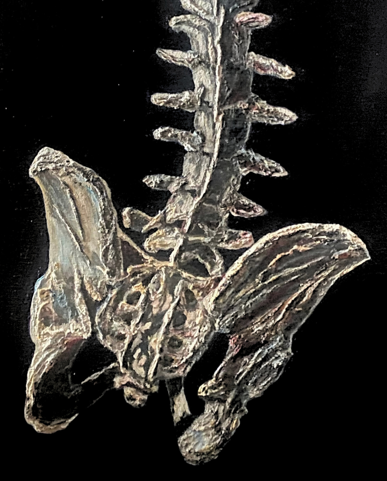
Intervertebral discs at some certain segmental levels undergo more degeneration than others which implies that mechanical stress plays a role in disc degeneration (8). The occurrence of dessicated or dried up discs mostly appear within the lower back or lumbar discs, in particular the lower three to four discs i.e. between L3-L4, L4-L5, L5-S1 and S1 (2).
However, any part of the spine can develop disc degeneration (11). As Griffith et al., (2007) highlight, Magnetic Resonance Imaging (MRI) is the most widely used method for assessing intervertebral disc degeneration. Based on proton density, water content, and chemical environment, an MRI depicts disc hydration and morphology (6).
How Serious Is It? Will I Need Surgery?
Within most cases, the majority of individuals diagnosed with the condition will not require surgery. Surgical intervention should be reserved for those with either neurological deficits, degenerative spondylolisthesis, or pain limiting daily functions (4) . Operative intervention of the lumbar spine include discectomy with fusion, total disc replacement or by immobilisation of the affected vertebrae (4). Surgery is offered only to one in every 2000 back pain episodes in the UK; the incidence of surgical treatment is five times higher in the USA (12).
Treatment
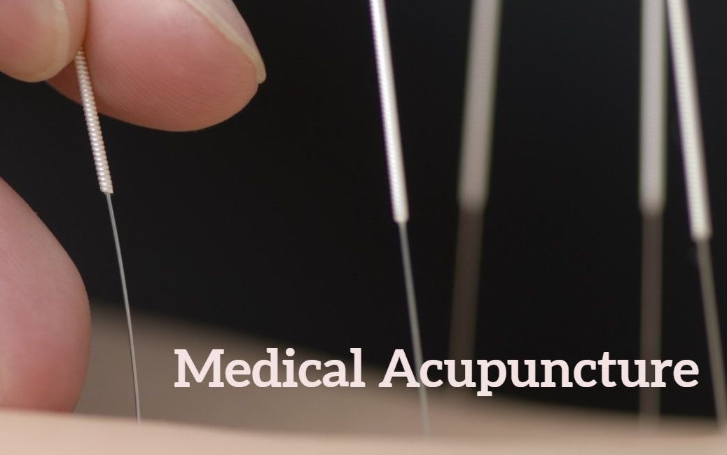
Therapies are designed to reduce pain and treatments are mainly conservative and palliative ranging from analgesia, corticosteroid injections, local anaesthetic and manipulation therapies all with the main aim of returning patients to work (13).
Physical therapy aim to strengthen neighbouring muscles around the affected joints before 'hands on' Spinal Manipulation Therapy (SMT)' can be considered as a treatment option, patients will need to be screened for possible serious pathology.
Osteopathy
Various physical therapy modalities might be applied. Osteopathic treatment may include gentle hands on joint mobility and articulation treatment techniques of the spine and surrounding tissues. The of use of hands on stretching and gentle joint motion mobilisation techniques might be applied along with exercise programs and electrical stimulation therapy such as interferential therapy, remedial massage techniques and modified lifestyle advise e.g. related to weight loss and smoking cessation.
Dynamic Lumbar Stabilization Program (DLSP)
Kessler
et al.,
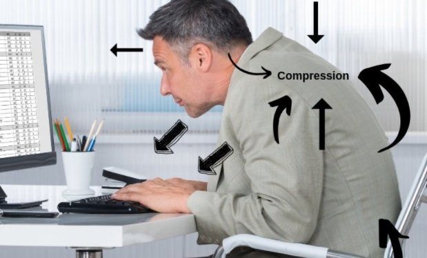
(2021) use of Dynamic Lumbar Stabilisation Program (DLSP) which aims to re-educate individuals' spinal posture. If observed and corrected early enough, reducing a lordotic lumbar curvature and neutralising the spine can improve body mechanics during every day living activities which can help slow down or lessen the degenerative process. According to their study, this has a high success rate (by up to 92-96%) with patients being able to return to work pain free. It also helps strengthen and retrain the deep multifidus muscle within the lumbar spine which helps spinal stability during movement and is associated with chronic lower back pain for if weak or atrophied (8).
The study by Faur
et al.,
(2019) found that the percentage of lumbar multifidus muscle atrophy is particulalry higher within the lower spinal levels L5 and S1. They concluded a low correlation and significant association between the grade of lumbar disc degeneration and the degree of lumbar multifidus muscle fatty atrophy. They discovered a higher fatty atrophy score in women and a greater incidence of disc degeneration in men (4).
Exercise Prescription
Exercises aims to improve the coordination between the abdominal and back muscles. Motion and strength training will help increase the strength and stability of muscles within the pelvic floor and for those that support the spine and pelvis. Stabilisation exercises increase the body's capacity to resist higher loads in the degenerative discs and reduce injury as muscular tissue reduces at a rate of 1 kg per year after the age of 40 and replaced by fatty tissue (1).
Training exercises performed one to three times per week reduces pain and should be continued once pain has subsided and patients are able to return to their jobs/hobbies or activities and are able to cease the use of analgesics (5). The Williams method involves stretching the back extensor muscles and strengthening abdominal muscles to relieve pressure placed on the lumbar intervertebral discs (5).
References
1. Alomari, R., Corso, J. J., Chaudhary, V. Dhillon, G. (2009) Dessiccation Diagnosis in Lumbar Discs From Clinical MRI with a Probabilistic Model, Research Gate, https://www.researchgate.net/publication/221624161_Desiccation_Diagnosis_in_Lumbar_Discs_from_Clinic... [online].
2. Brzuszkiewicz-Kuzmicka, G., Szczegielniak, J., Baczkowicz. (2018) Age
related changes in shock absorption capacity of the human spinal column, 13: 987-993.
3. Donnally, C. J., Hanna, A., Varacallo, M. (2023)
Lumbar Degenerative Disk Disease, StatPearls Publishing LLC [Online], Last visited 12/01/2024.
4. Faur, C., Patrascu, J. M., Haragus, H., Anglitoiu, B. (2019) Correlation Between Multifidus Fatty Atrophy and Lumbar Disc Degeneration in Low Back Pain, BMC Musculoskeletal Disorders, 20; 414: 1-6.
5. Gąsiorowski, A., Zagórski. J. (2013) Strength training in the treatment of degeneration of lumbar section of vertebral column, Annals of Agricultural and Environmental Medicine, Vol 20; 2: 203–205.
6. Griffith, J. F., Wang, Y-X. J., Antonio, G. E., Choi, K. C., Yu, A., Abuja, A. T., Leung, P. C. (2007) Modified Pfirrmann Grading System for Lumbar Intervertebral Disc Degeneration, Spine, 32; 24: pp E708 –E712.
7. Hawkins, J. L., Denson, J. E., Miley, D. R., Durham, P. L. (2018) Nicotine Stimulates Expression of Proteins Implicated In Peripheral and Central Sensitisation, Neuroscience PMC, 290: 115-125.
8. Kessler, R., Haase, C. Dean, D. (2021) An Osteopathic Approach to Patients with Degenerative and Herniated Discs, The AAO Journal, 31; 2: 35-41.
9. Kos, N., Gradisnik, L., Velnar, R. (2019) A Brief Review of the Degenerative Intervertebral Disc Disease, 73; 6: 421-424.
10. Lee, B. H., Moon, S-H., Suk, K-S., Kim, H-S., Yang, J-H., Lee, H-M. (2020) Lumbar Spinal Stenosis: Pathophysiology and Treatment Principle: A Narrative Review, Asian Spine, 14; 5: 682-693.
11. Medline Plus (2016) Medline Plus: Trusted Health Information for You, National Library of Medicine, Degenerative Disc Disease, [Online]
https://medlineplus.gov/download/genetics/condition/intervertebral-disc-disease.pdf last visited 11/01/2024.
12. Meyer, F.,
Börm, W.,
Thomé, C. (2008) Degenerative Cervical Spinal Stenosis, 105 (20): 366–372.
13. Urban, J. P. G., Roberts, S. (2003) Degeneration of the Intervertebral Disc, Arthritis Res Ther, 5; 120.
14. Xin, J., Wang. Y., Zheng, Z., Wang, S., Na, S., Zhang, S. (2022) Treatment of Intervertebral Disc Degeneration, 14: 1271–1280.
15. Yanagishishita, M. (1993) Function of Proteoglycans in the Extracellular Matrix, 43; 6: 283-293.