Buttock Pain & Piriformis Syndrome
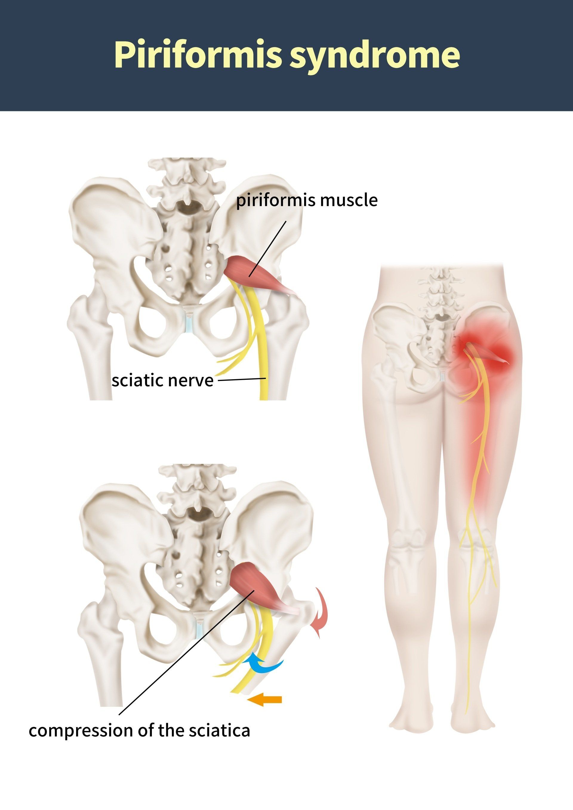
Piriformis syndrome is a painful musculoskeletal condition that resembles sciatica and secondary to sciatic nerve entrapment within the piriformis muscle at the greater sciatic notch (2).
Cadaver dissections gave Beaton and Anson (1938) the hypothesis that muscle spasms of the piriformis muscle could be responsible for the irritation of the sciatic nerve.
The term 'piriformis syndrome', however, was first introduced by Robinson (1947)who described that the cardinal features (main clinical signs) of the syndrome have six criteria (1).
- A history of trauma to the sacroiliac and gluteal regions.
- Pain within the region of the sacrolilac joint, greater sciatic notch and piriformis muscle which may extend down the leg and cause difficulty walking.
- An acute exacerbation of pain caused by stooping or lifting.
- A palpable sausage shaped mass (or muscle knot) which is tender to palpate over the piriformis muscle on the affected side.
- A positive Lasegue sign (viewed below).
- Gluteal atrophy.
Piriformis Syndrome commonly occurs from abnormalities of the piriformis muscle such as (2):
- Hypertrophy.
- Inflammation.
- Sciatic nerve irritation or entrapment.
- Lumbar radiculopathy
- Sacroilitis
- Trochanteric bursitis
- Intervertebral discitis
- Trauma
- Excessive exercise
- Leg length discrepency
- Altered Biomechanics
- Narrowed sciatic foramen
- 'Pocket or wallet sciatica' caused from sitting on wallets in the back pockets of trousers or jeans.
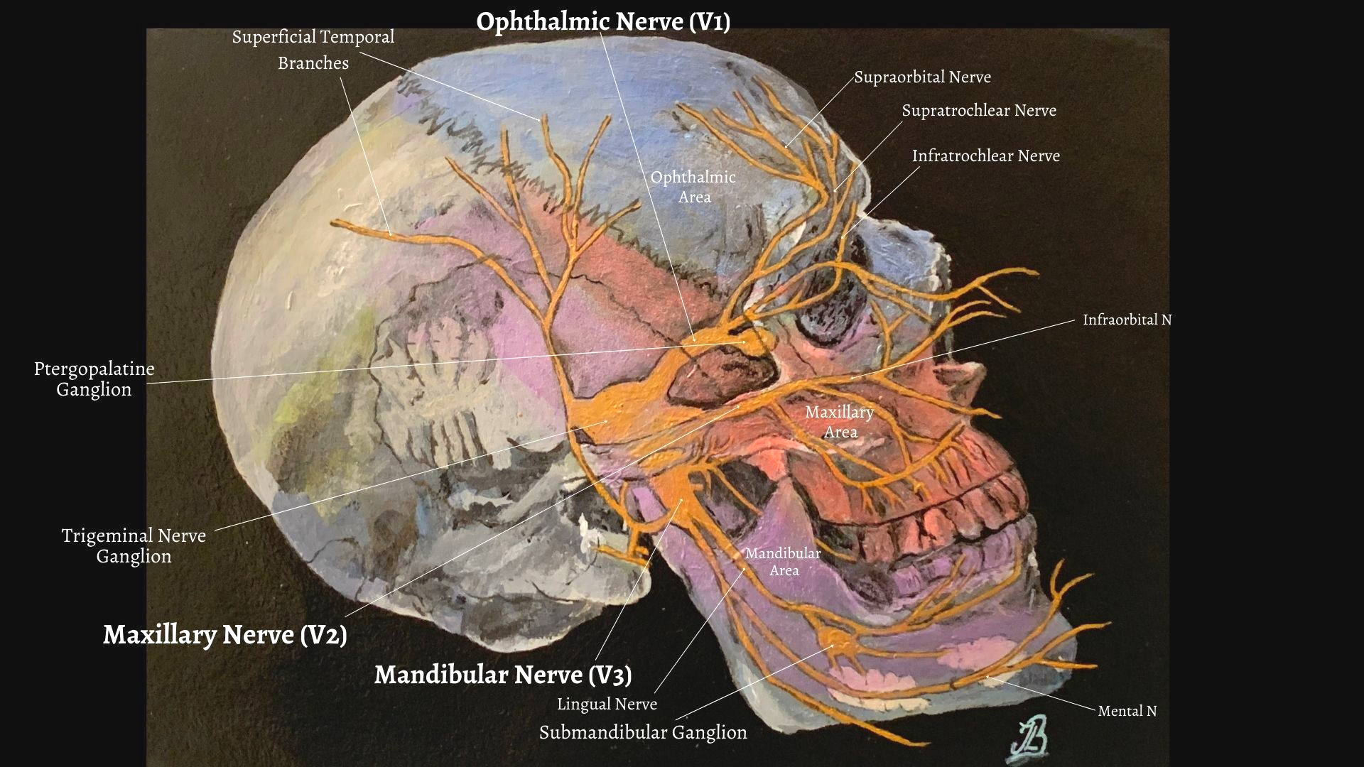
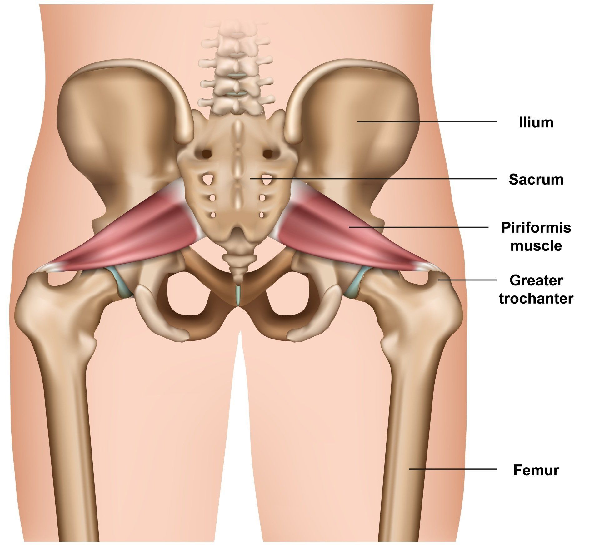
Anatomy of the Piriformis Muscle:
Origin:
The piriformis muscle originates from the anterior surface of the sacrum (S2 vertebrae to S4), upper margin of the sciatic nothch, the sacrotuberous ligament and adjoining areas of the sacroiliac joint.
Insertion:
The piriformis then inserts into the superior medial aspect of the greater trochanter of the femur and the tendon often joins with the tendons of other hip external rotators; the superior gemellus, inferior gemellus, and obturator internus muscles prior to its insertion (2).
There are overall 6 external rotator muscles of the hip which are in close proximity to one another and work as a functional unit:
- Piriformis
- Superior gemellus
- Obturator internus
- Inferior gemellus
- Obturator internus
- Obturator Externus
- Quadratous Femoris
Diagnostic Testing:
There are several clinical tests but no single test is specific for piriformis
syndrome (2).
- Piriformis sign - In supine position when the patient is relaxed the ipsilateral foot is externally rotated and active efforts to bring the foot in midline results in pain which is otherwise a positive sign.
- Lasegue sign (or straight leg raise (SLR) test) - In a supine position, the hip and knee is flexed to 90 degrees, then keeping the hip flexed extend the knee , if the patient reports posterior thigh pain it is a positive lasegue test.
- Freiberg sign - Pain is experienced during passive internal rotation of hip joint.
- Pace sign - Pace sign, revealed with the FAIR (flexion, adduction, and internal rotation) test, involves the recreation of sciatic symptoms.
- The FAIR test - performed with the patient in a side lying position, with the affected side up, the hip flexed to an angle of 60 degrees, and the knee flexed to an angle of 60 degrees to 90 degrees. The hip is stabilised and the examiner internally rotates and adducts the hip by applying downward pressure to the knee. Alternatively, the FAIR test can be performed with the patient supine lying or seated with the knee and hip flexed, the hip is medially rotated. The patient resists external rotation and abduction of the hip. The FAIR test result is positive if sciatic symptoms are recreated.
- Beatty test - Lying on the unaffected side, lift and hold the superior knee approximately 4 inches off the examination table. If sciatic symptoms are recreated, the test result is positive.
Other External Rotator Muscles of the Hip
The external rotator muscles of the hip are in close proximity to one another and work as a functional unit.
- Piriformis
- Superior gemellus
- Obturator internus
- Inferior gemellus
- Obturator internus
- Obturator Externus
- Quadratous Femoris
Treatment for Piriformis Syndrome
Manipulative treatment of piriformis syndrome would mainly have the aims to restore normal range of movement and reduce pain by reducing the tightness of the piriformis muscle. Techniques used to acheive this goal may include techniques such as muscle energy techniques, joint articulation, high velocity/ low amplitude manipulative therapy treatment, massage, motion controlled and strengthening exercises, stretching techniques and exercises. Other studies suggest that additional physiotherapy modalities that may also help are heat therapy, cold therapy and sonopheresis or ultrasound (2).
References:
1.) Jawish, R. M., Assoum, H. A., Khamis, C. F. (2010) Anatomical, Clinical and Electrical Observations in Piriformis Syndrome, Journal of Orthopedic Surgery and Research, 5; 3: 1-7.
2.) Mitra, S. R., Roy. S. Dutta, A. S., Ghosh, A. Roys, R. Jha, A. K. (2014) Piriformis Syndrome: Review Article, J or Evolution of Med and Dent Sci, 3; 14: 3804-3814.